Fruitflow®: What Is It and What Are Its Health Benefits?

Fruitflow® is a natural extract from the jelly around the seeds of sun-ripened tomatoes that has been found to have beneficial effects on platelet aggregation and cardiovascular health.
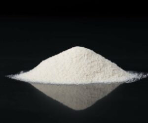
It is a water-soluble tomato concentrate that helps maintain normal platelet aggregation, …